- >>
- Healthy Adult Studies
- HCP Young Adult
- News
- Article
Electrophysiological origins of connectivity: Characterizing resting state networks with MEG
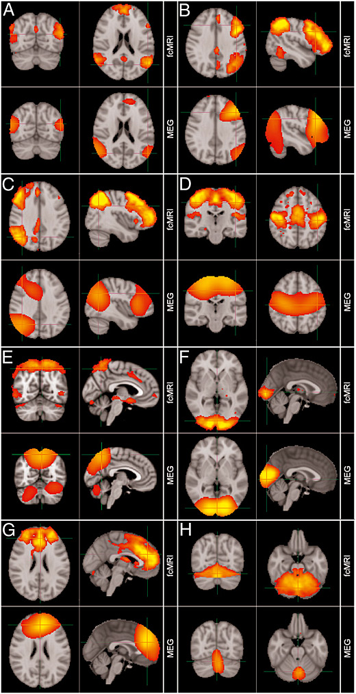
A new paper published in PNAS by HCP investigators and their MEG collaborators in the United Kingdom uses magnetoencephalography (MEG) to independently investigate and characterize Resting State Networks (RSNs) in the human brain. MEG offers a useful way to measure connectivity between brain regions because it bypasses the hemodynamic BOLD response (an indirect measure of brain activity used in fMRI) and measures the electrophysiological basis of brain activity.
The authors describe a means to assess connectivity in MEG data by a unique combination of beamformer spatial filtering and independent component analysis (ICA). Their results have strong correlation with the spatial structure of RSNs that had been previously identified with fMRI, and confirm the electrophysiological basis of hemodynamic networks in the brain. The work also demonstrates the potential of using MEG as a tool for studying brain networks.

The paper was authored by Matthew J. Brookes and Peter G. Morris of the University of Nottingham and co-authored in part by HCP investigators Mark Woolrich and Stephen Smith of Oxford University.
PNAS.org: Investigating the electrophysiological basis of resting state networks using magnetoencephalography